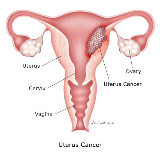
Management of all types of Uterus Cancer
Uterus or "womb" is the female reproductive organ where the foetus develops. Abnormal growth of cells in uterus is called as uterus cancer.
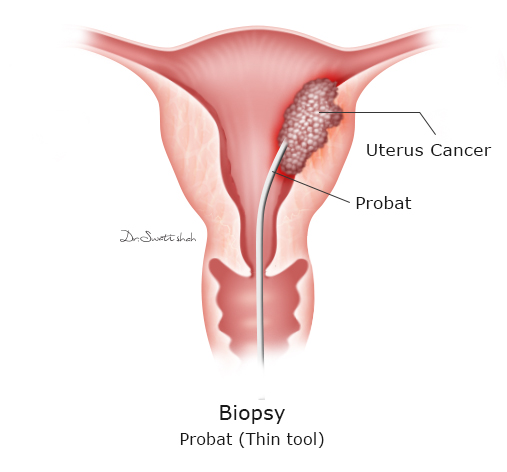
Tissue samples from the uterus are taken out and checked under microscope. This tests finds out if there is cancer in uterus or not.
A thin & flexible tube is inserted into the uterus to take tissue samples to confirm if there is uterus cancer or not.
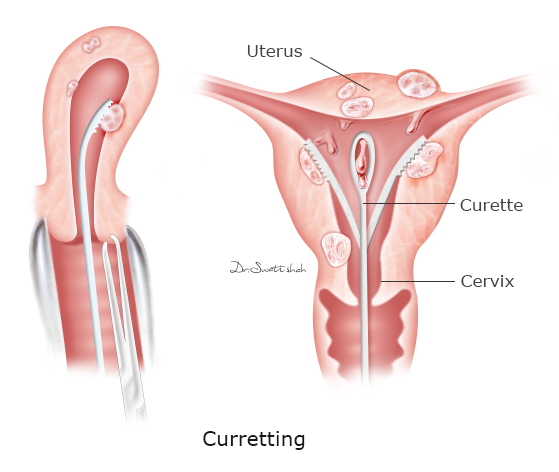
Tissue samples are taken from the uterus with help of a curette after dilating it. This procedure is done under anaesthesia.
Sonography is done through vagina to have a better look at the uterus and ovaries. It will identify if there is any suspicious area or tumour in uterus or not.
After diagnosis and staging with MRI/ CT scan or PET scan, surgery is the 1st step in treatment of Uterus Cancer in most patients. Surgery for Uterus Cancer includes :
Uterus is removed along with cervix, both ovaries, upper vagina and tissues surrounding it. This surgery is quite different from Simple Hysterectomy done for non cancer conditions. In Radical Hysterectomy, removal of extra tissues help to remove uterus cancer completely.
This is important step in Surgery for Uterus Cancer. Cancer cells can spread from Uterus to Lymph nodes in adjacent areas (Pelvic) and these nodes have to be removed.
Uterus cancer can spread to nodes along major vessels and these are also removed.
Uterus Cancer Surgery is best done by Robotic Surgery.
Uterus Cancer Surgery can be done by Open / Laparoscopic or Robotic approach. The surgical procedure is same in all three, but the approach is different.
Surgery is done though vertical cut on abdomen. Patient requires 5 to 7 days of hospitalisation and around 15 days for recovery.
Surgery is done through small holes on the abdomen. Camera and instruments are inserted through these holes and complete procedure is performed. Patient needs to stay in hospital for 2 to 3 days only and recovers very fast.
It is same as Laparoscopic Surgery in approach (through small holes), stay & recovery with some additional benefits. Better View ( 3D, 10X magnified & HD View) and better instruments (flexible) make the surgery more precise and accurate. Less blood loss, less pain, better nerve preservation enhances functional recovery of the patient.